Description
This video was filmed and produced by Gil Hedley. Please note that It includes graphic videos and photos of dissections of cadavers (embalmed human donors). You can visit his website for more information about his workshops.
About This Video
Transcript
Read Full Transcript
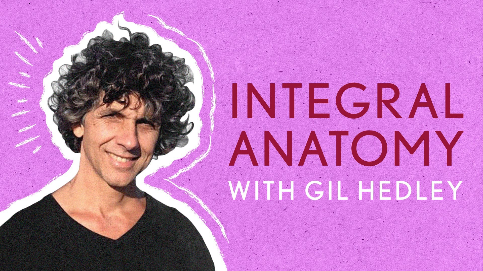
Greetings and welcome to the Integral Anatomy Series. My name is Gil Headley, and I'm grateful for your choice to join me in this exploration of human form as Psalmonauts dedicated to exploring inner space. Images and information briefly reviewed in the first few minutes of this volume are given the full attention they deserve in the first volume of this series. Please include that first volume in your own set of learning experiences to provide the full context and foundation for your understanding of deep fascia and muscle. As we move through the following sequences, we'll see the elegant grid pattern, which arises here from the confluence of wind and wave reiterated in the relative stillness of the human form.
Fluid movement leaves its signature tracks upon all manner of materials on our planet, imprinting common patterns in space upon substrates with vastly different relationships to time. Our study of the cadaver forms amounts to the viewing of still snapshots and artifacts of the life lived and patterns traced in the living tissues. Those patterns ultimately yield to the flow of matter in the larger cycles of movement in our universe, and we remember and acknowledge with gratitude those who have passed. We thank the families of the donors, and we attend carefully to the gifts they have left us. Yet through the ever original combination of matter, motion, human intent, and the intelligence of nature, new forms will be manifest to enrich our life experience as we pursue these ongoing experiments with balance, relationships, and a connection to the hidden layers of ourselves.
The dissection progression represents a step-by-step movement into the deeper levels of the human form. In this series, we study the body at the macro level. Here we intend a deep learning experience with the level of physical form which can be readily observed with our eyes, felt with our hands, differentiated with a fingertip or scalpel, and then reflected and removed for further appreciation and study. The amazing worlds of microbiology, molecular biology, and physics remain outside the range of the camera lenses used on this particular project. Because patterns of relationship and structure have been proven to repeat at many different levels, observations made at the gross level give us a very practical experience of the kinds of relationships which exist at other important levels of connection within us and between us.
Applying the onion-tree model to the whole body is a lens which focuses our attention particularly on layered shifts of tissue textures and head-to-toe continuities which the human form presents. There are an infinite number of other ways one could choose to dissect a body, and each one would reveal something new, something previously overlooked. I have experienced this layered approach to be a very powerful learning tool without needing for it to be the only word or the last word on the subject. There really are no layers in the integral human form. That having been said, look at this amazing layer.
When I speak of deep fascia in this presentation, I am referring very specifically to the mostly fibrous though sometimes filmy, sometimes translucent and often opaque and whitish layer that my hands and scalpel come up against just deep to the relatively soft and loose areolar superficial fascia and before coming to the red-brown muscle fibers. The relationship of the deep fascia to the superficial fascia was explored in volume 1. Trying to understand deep fascia without studying skin and superficial fascia is like trying to understand a marriage while ignoring the spouses. So here we are observing the varied surface of the deep fascia in place as it first presents to us before we explore the underlying relationship of the deep fascia to the very different texture of the muscle. Despite the stillness of the cadaver, the impression of fluid movements, patterns and energetic pathways are evident in the fascia.
Every body is in some sense a physical hologram of interfacing waveforms of varying vibrations. Deep fascia falls into the much broader category of connective tissue. All fascia is connective tissue, but not all connective tissue is fascia. Connective tissue also includes blood and bone, the perineural and perivascular wrappings and the entire omnipresent web that invests, surrounds and interpenetrates every cell in stunning continuity right down into the cytoskeletal microtubules. Researcher James Oschman directs our attention to the spectacular whole body communication network which the connective tissue matrix represents.
Robert Becker demonstrates it as a locus of healing and energetic phenomena. Robert Schleip and his colleagues at fasciaresearch.com have definitively demonstrated the active contractility of the fascia, which is in fact replete with specialized smooth muscle fibers. Not merely the skeletal muscle, but the fascia itself also actively contracts. Fascia is responsive and alive. I point out the anatomy of the fascia at a gross physical level, revealing its context and continuity across conventional regions.
While skin covers the surface of the body, deep fascia covers the muscle layer, but it also spans the depth of muscle and relates through its many fibrous septae right down to the bone. I habitually use the metaphor of a bag when I dissect deep fascia. The deep fascia is no more a bag than is a cell membrane, since the idea of a bag may evoke a sheet-like quality, and deep fascias are related through countless vectors by fuzzy, filmy, fibrous, vascular, neural, and perineural trees, just as cell membranes are interpenetrated by the flexible cytoskeleton, linking with the extracellular connective tissue matrix. Nevertheless, anatomy generates abstractions by the pound. My use of the bag metaphor for deep fascia speaks directly to the ease with which I can cut it up and make it appear as a sheet.
I create the bags in order to define and clarify different tissue textures. In the animate, living human form, deep fascia, like all the other layers of the body, is comprised of more than just fibrous cells and sundries. It is shot through with the pulsing, vibratory phenomena of life itself, precisely what is absent here from the cadaver form. When you take some time to internalize your experience of this program, be sure to notice and feel the joy of life expressed in the great gift of your human form. This is a famous fascia.
It's called a fascia lata. It's a particular name given to an area of the deep fascia, which covers the whole body. We have a bag that covers the whole body, and then we have particular names for particular areas of the fascia. Famous fascias, you could call them. This is a famous fascia.
It's called a fascia lata. It's this fibrous bag that envelops the whole thigh and, of course, is in continuity with the rest of the deep fascia of the body. We'll focus in maybe around here, and it'll allow the viewer to see some of the thin, filmy tissue above the deep fascia, which disguises its fibrous nature. What we're looking at are remnants of the superficial fascia, but not merely remnants. We're looking at the relationship, the remaining relationship of the superficial fascia to the deep fascia.
Let's get even a little closer here. Let's see how close we can get to some of this tissue right here. We'll see that there's something of a film. Here we have this beautiful, strong fibers here coming in this direction. On top of it is something of a film.
If I can lift up this kind of filmy tissue here, and we can see... Okay. When you see those beautiful pictures in the Color Atlas, they've gotten all of this film off. It's the most tedious process. It's fun, but it takes a lot of time and patience, and while you're doing it, you often poke holes in the bag underneath, and then it doesn't look as good for the picture. I'm more concerned in this instance to demonstrate what it takes to get the picture, and then you have a sense, a little more of the reality of the cadaver form and our living tissues as well.
I back off a little bit of this tissue, and we can see... I can scrape a little bit with my scalpel. I'll try and keep my shadow out of here, and I'm lifting this film away, and then we see more of that stripey quality, which underlies it. We have a form that's approximately six feet tall. The deep fascia covers the whole body, so rather than spending the next month, which I could easily do, perfecting this image, I'm trying to give you the concept of this beautiful stripeity strapping that constitutes the textures and depth of the bag of the deep fascia as it overlays the musculature of the thigh. We have a beautiful multi-directional pattern here.
Right here, this little dot here is a perforating vessel that's been severed. This would be blood flow to the superficial fascia. The trees are perforating the bags. I think it would be most instructive to actually go inside the bag. Let's see if we can capture the whole thigh here at the same time, and give a sense of what it'll mean to enter this space.
If I lift up right around here over the knee, I can tug at the bag, and I can give a little tug all along here, and start to differentiate the bag of the deep fascia from the tissue that's underneath it. By differentiating it first, that enables me to spare myself an untoward cut into the musculature. I can actually get my pinky under here a little bit, and start to get air. I don't know if it's possible to see that air right now. Let's see.
You can see the air creeping in under the bag now. I get my hemostat under here, and I can start to probe my way along this pathway. I'm neither poking into the muscles, nor am I making holes in the deep fascia. I place my hemostat inside the bag. It's pretty cool.
So you've got a sense of the depth of the bag. It's transparent. I can see the silver of the hemostat through the bag. It's not a thick bag, and yet it's a bag with tremendous integrity. Now relating the deep fascia to the musculature itself, even in places where it's easy to slip and slide the deep fascia off, there's sort of fuzzy connective tissue relationships that are more or less present in a given form.
Ultimately, though, this is a fairly light relationship. The rectus femoris, which underlies the tissue here, can ultimately slide in this relationship with the fascia. So I'm actually going to incise this now with my scalp, and now I know where I'm going. I'll cut it, let's say, along the line of the rectus femoris here. As I use my scalpel, I lift with my hemostat and lift the bag so I know that I'm just cutting the bag.
I can even slip my scalpel underneath and lift it. I'll bring my probe under some more so that I make sure I'm differentiating the tissues that I want to. And ultimately, we'll be able to, just like we did with the skin in the first DVD, and just like we did with the superficial fascia. Here's an opportunity that we'll have to see through the deep fascia, to understand the deep fascia in its depth and integrity as a whole body layer, an organized, a highly organized layer, and very beautiful. What we have here is this fuzzy relationship of the deep fascia to the muscle below.
This is really pretty. And the light is picking up nicely on the fibers, and as I lift even more, I'm actually lifting the epimesium off of some of the muscle fibers below as I yank on the deep fascia. So you get a real sense here, I hope, of the intimate relationship of the deep fascia to the tissue beneath it, and that when I create a singular plane out of the deep fascia, I'm doing so as an anatomist, as someone creating abstract realities from physical realities. Because the tissue, in fact, is not a plane. It's multi-dimensional, multi-directional.
And it's got the muscle in the bottom there. Well these are muscle fibers here. And this is the deep fascia here. And this here is the relationship of the epimesium, the fascia around the muscle cells as they enter into a relationship with the deep fascia. So the deep fascia is a different texture altogether, and yet it's not like it's not connected.
See everything is connected, but what happens is you get these transitional textures from one layer to the next. And that's what studying relationships is all about. In a sense, as I yank up on the otherwise flat layer of the epimesium, you're able to see that it's structure is very light and diaphanous and fluffy. And keep lifting this bag, here's a blood vessel, remember we saw a little something coming up through on this side, remember that tiny little blood vessel we talked about? Well when we lift the bag, we look on the other side and we see it in its length coming here, a little vein, I'm actually going to cut it.
The marriage of the layers often happens through perforating vasculature and nerves, etc. It looks like a sheet when I lift it up, but it's actually a curve, just like the surest distance between two points on the surface of the globe is an arc, not a straight line. All of the tissues of our body are arc tissues. And we see fatty tissue here. So fatty tissue isn't only limited to superficial fascia, there's also fatty tissue intervening at transition points from one muscle bag to another.
Our body has no empty places in it, so if there's a kind of a wedge or something to be filled in, it'll be filled in with adipose tissue, loose aerial or adipose tissue. So I'm going to scratch my way along here and see if I can't get underneath this transition point where the tensor fascia lata is embedded in the fascia lata itself, in the deep fascia and acting upon it. I can actually take my finger, trace it through this fuzz, and the fuzz just melts to my finger. It's not tough at all, reflecting the deep fascia, and we see it from a different perspective, and it's possible to sort of peek in at what I'm looking at on this side of the table here. And if I press here and slide with my fingertip, I can kind of clean the tissue off and in a sense free the tissues that have always been intimately bound.
Now ultimately you would think at this rate I could be able to go around the whole form from one side of the thigh to the other and back around, but in fact what's going to happen is we come to the end, it's a compartment, there's a stopping point where the tissue of the deep fascia dives under to the bone. It's on the surface and then an inner portion of it dives under to the bone and connects to the bone, creating a compartment for this musculature relative to the musculature of the hamstrings. Or in the front over here we'll run into what's called a septum, where the hand can slide no more in the bag because the bag itself sends down, scrolls its way down, creating a particular compartment for these muscle tissues here relative to other muscle tissues. See that grid? It's incredible.
So the grid is going in both directions. Okay, now let's hold it aloft a little bit, oh nice, and then we'll sneak underneath it and we'll see the tons of fascialata and the various reds, etc. There you can see the vertical on the right side as well. Of course as always I'm disrupting the deep relationship of the deep fascia to the musculature underlying it, which is sometimes a fuzzy relationship, sometimes a fibrous relationship, sometimes a slippery, slidey relationship, and it would be a very worthwhile, complex and enduring study to identify completely for the entire body exactly what is the normal relationship of the deep fascia to the underlying layer in any given place. See here it's quite the bag, quite the fibrous bag just like the fascialata, and still we see the muscle fibers in relationship with it until I scrape them back and then they seem to have an integrity of their own and yet I think if we put a microscope to it we'd see that I've cut the attachments of the muscle fibers here, that's what it feels like, I'm scraping the muscle fibers away from the deep fascia, see I can take my scalpel and drag it along the shiny fascia as I attempt to break through the septum from one muscle balloon to the next, which we can see here, the septum.
Now at this point I'm sneaking up on the retinaculum of the wrist, it's stronger and strappier here and it's thin again here, the same tissue covering and yet it's just a bit more fibrous here. So these photographs in the atlases and the drawings by the artists representing the retinaculum, if you want to create that sort of thing you just, well you cut here and you could do the same up above, if you really want to make a retinaculum, famous one, let's do it, let's create the extensor retinaculum, see I pull back the deep fascia and slice it away from its relationship here, I create a border and if I just kept peeling all this away on the hand and lifting it up from its relationships and sawing the tissue back, it's like I'm removing a latex glove almost, so I pull this bag away and if I leave this here I can trim it even more until only the shiny silver fibers are there and then you take a picture and you say that's the, or you draw a painting and you say that's the extensor retinaculum and it is. If you look very closely in between each muscle fiber, it's sort of a yellow line or a whitish line depending upon the lighting here, what that represents is at this point the cut of my knife differentiating the deep fascia off of the muscle and it's as if at this point analogous to where in the leg when I differentiated the fascia lata and it threw off those giant septae between the muscle bundles, here we have the deep fascia throwing off septae but between muscle fibers and so we end up with a striped look because at these points along these lines in between these muscle fibers we have a rising up connective tissue that's rising up to meet the deep fascia that I'm peeling back, so it's not as if this deep fascia that I'm, this plane of tissue here has any substantive reality other than as an abstraction from the cutting of my knife I pull back on the tissue and my scalpel goes dull severing the deep fibrous connection from the covering from the fibrous covering so the covering is only a covering to the extent that I cut it away from it's third dimensional roots, there are no independent structures in human form, the human form it may even be a mistake to consider the human form itself as an independent structure because it's embedded in its environment in ways that we cannot necessarily perceive with our eyes and yet there are relationships, vibrational relationships between everything in a given environment and those relations strike me as no less or more significant than these physical connections that I'm demonstrating here as I peel back deep fascia with my scalpel and hemostat where does one tissue end and another begin well pretty much wherever we decide because to the extent that we create division or identify one thing from another in the human form we are basically writing a mythology or creating a model that at some point must be dumped for a deeper understanding if we cling to a model that may have worked for a while it's like refusing to go further in the learning process I speak of several layers of the form as a way of augmenting the regional anatomical model and creating yet another way of looking at the body however it's also true that my model here of the layered form or the onion tree with layers and interstitched with tree like structures the vasculature of the nerves will also develop and become more sophisticated we mustn't cling to our models too tightly. Thank you.
Integral Anatomy: The Integral Anatomy Series
Comments
You need to be a subscriber to post a comment.
Please Log In or Create an Account to start your free trial.


