Description
About This Video
Transcript
Read Full Transcript
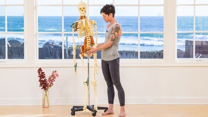
All right, welcome back. So in this segment, we'll look briefly at the psoas muscle, which I talked about quite a bit in one of the practices, and we'll also look at the hip joint. And I'd just like to point out which parts of the hip joint I might have referenced. So to begin with, we'll talk about the psoas, and this anatomical model has a psoas. And the psoas is the only muscle in the body that literally connects your axial body, that's your spine and torso, to the lower appendage, your leg.
Here you can see that the psoas starts just below your bottom ribs. And at the bottom ribs, there's another sheet of muscle. It attaches all the way along the circumference of the ribs. It's called your diaphragm, otherwise known as your breathing muscle. And the fibers of the psoas are continuous with the diaphragm.
So where the psoas starts is right below your bottom ribs at the top of your lumbar spine. And it is attached to the sides of your vertebral bodies. And in just a minute, I'll turn the skeleton. You can see how far forwards the front of your spine is. But I'll say here that the psoas is attached to more of the front part, or the side of the front of your spine.
So it attaches along the length of your vertebral bodies, down to the last lumbar vertebra. And then here, it teams up with another muscle called the iliacus. And the iliacus is this muscle here that lines the inside of your hip bone. Now when they team up, they're called the iliopsoas. And they both then attach to what's called the lesser trochanter.
So the lesser trochanter is this little knob, this kind of sticky, outy part on the upper inner thigh bone, right? So that's the psoas muscle. Now when we look at the hip joint, the psoas runs across the front of the hip joint. And there's a whole bunch of other stuff, attachments and connective tissue, that also cross the front of the hip joint. But here you can see that the ball of the femur, that's your thigh bone, is in the socket of the acetabulum.
And I'm going to turn this model to the side, right? So the hip is a ball and socket joint. The socket or the acetabulum is very deep. The head of the femur, that's the ball part, sits deep within the socket. And if you find your own belly button and go out to the side, about two or three inches, that's about where your hip joint is.
So a lot of us tend to think of our hips as more to the outside. But in reality, the hip joint is fairly deep in. And this bony protrusion or this knob right here is called your greater trochanter. So the inside, you have the lesser trochanter. And then the outside, you have the greater trochanter.
And the greater trochanter is a part of your anatomy that is possible to feel. So if you feel the top of your thigh bone, you'll probably be able to palpate your greater trochanter. So this, when I talk about the hips, I would talk about this as the outer hip. Now continuing on, we turn the skeleton to the back. So I'm going to pull this back.
So here you can see that the lesser trochanter is a little bit further situated to the back. And it's close to your sit bone. So when I was talking about the psoas, and I said that you can feel the lesser trochanter slightly diagonal and down from the sit bone, that's the relationship right there. So again, the psoas here would attach from the front onto this knob right here. And then this portion is the back of your hip joint.
So when I talk about the hips, this would be the back portion of the hip joint. It's between your sit bone and your greater trochanter. That's where the joint capsule is. All right. So I hope that clarifies some of the anatomy for you.
And it's always great just to look at models or look at pictures and then do your own self-touch and palpation to figure out where things are. Learning about anatomy can be fun, experiential, and really helpful when you're moving. Thanks for joining.
Anatomy for Yogis: Renee Sills
Comments
You need to be a subscriber to post a comment.
Please Log In or Create an Account to start your free trial.


